Glaucoma is usually associated with the build-up of fluid pressure inside the eye. If left untreated it can cause vision loss by damaging the optic nerve (the wire that connects the eye to the brain).
Glaucoma is commonly referred to as “the silent thief of sight,” because in most cases it progresses gradually and quietly. Vision can be damaged without any noticeable symptoms. It may begin with loss of peripheral vision and then progress to central vision loss and blindness. It affects nearly two out of every 100 people over the age of 35. Half of those are at risk for blindness because they may not even know they have the disease. At Bennett & Bloom Eye Centers, our doctors use the most up-to-date technology to diagnose and treat glaucoma. With early detection and careful management, significant damage to eyesight from glaucoma is nearly always preventable, although treatment cannot restore vision that has already been lost.
What is glaucoma?
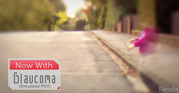
Glaucoma is a complex disease associated with the build-up of fluid pressure inside the eye that can damage the optic nerve. The optic nerve, a bundle of over a million nerve fibers, transmits the message of sight from the eye to the brain. In glaucoma, the nerve fibers carrying peripheral vision are affected first. This reduction in side vision can be gradual and is usually asymptomatic. By the time it affects central or reading vision, tremendous damage to the nerve has already occurred.
 Normal vision

Glaucoma causes side vision loss that can often be completely asymptomatic
To understand what’s happening with glaucoma, imagine the eye as a sink with water. The clear fluid inside the eye (aqueous humor, produced by the ciliary body) and is always flowing into and out of the eye, just like a sink with an open faucet and drain. As long as the drain is open, a sink won’t overflow. But if anything happens to block the drain, the water level rises and spills over the edge. Since the eye is a closed ball, blockage to its drain (the trabecular meshwork) and pipe (Schlemm’s canal) doesn’t cause aqueous to overflow or leak out. It has nowhere to overflow. Rather, the eye’s fluid pressure increases and damages the optic nerve.
What are the types of glaucoma?
Although there are many different causes for glaucoma, there are two broad categories: open angle and narrow-angle. The most common type is the chronic open angle glaucoma. In this condition, the opening into the trabecular meshwork is working but is internally clogged, causing the pressure to painlessly and gradually increase. Vision is lost gradually in the periphery so that it is generally not noticed until significant damage has occurred.
The other major type of glaucoma is narrow (closed) angle glaucoma. With narrow angles the iris (the colored part of the eye) blocks the trabecular meshwork’s opening, causing a sudden severe rise in eye pressure. This can cause halos around lights, severe pain, and rapid vision loss.
Both of these types of glaucoma can be inherited. If someone in your family has glaucoma, your risk of developing it is increased. Likewise, if you have glaucoma, others in your family need to be made aware that their risk is increased. There are numerous other types of glaucoma, including secondary, juvenile, pseudoexfoliation syndrome, and pigment dispersion syndrome.
How is glaucoma diagnosed?
At Bennett & Bloom Eye Centers, our doctors use the very latest technology to identify patients with glaucoma. The diagnosis is not always clear-cut since each eye varies in its susceptibility to eye pressure. Successful diagnosis and treatment begin with a careful ocular evaluation. First, we measure the fluid pressure within the eye. Corneal thickness may affect this pressure reading and can be checked with an ultrasonic pachymeter. We examine your eyes thoroughly and look for optic nerve damage, both visually and photographically. Using our state-of-the-art OCT we can measure the nerve fiber layer thickness around your optic nerve. This test may determine if you have glaucoma even before you experience any vision loss. Next, we test for central and peripheral vision loss using a sophisticated, computerized visual field analyzer. Gonioscopy helps determine which specific type of glaucoma you may have.
We’re always careful to consider any additional risk factors including family history, general health problems, including diabetes, anemia and prior eye trauma.
Finally, we analyze all the information to arrive at your unique diagnosis and treatment plan if indicated. Usually, prescription eye drops are the only treatment needed to prevent further vision loss. More advanced cases may require a laser or surgical procedure.
How is glaucoma treated?
- Open-angle glaucoma
- Eye drops
In most cases, open-angle glaucoma can be treated with eye drops taken once or twice daily. These drops help lower eye pressure and reduce the risk of further vision loss. Sometimes more than one drop is needed. If drops do not adequately control eye pressure, laser or surgery may be needed. There are many types of glaucoma drops:- Alpha agonists: Reduces the production of fluid in the eye. Drugs include Alphagan P (brimonidine tartrate) and Iopidine (apraclonidine hydrochloride).
- Beta-blockers: Reduces the production of fluid in the eye. Drugs include Betimol (timolol), Timoptic XE (timolol maleate), and Betoptic S (betaxolol hydrochloride).
- Prostaglandin/prostamide analogues: Improves the outflow of fluid. Drugs include Travatan (travoprost), Lumigan (bimatoprost) and Xalatan (latanoprost).
- Carbonic anhydrase inhibitors: Reduces the production of fluid in the eye. Drugs include Trusopt (dorzolamide) and Azopt (brinzolamide)
- Cholinergic agonists (Pilocarpine): Improves the outflow of fluid from the eye.
- Multi-mechanism agents: Drugs include Rhopressa (netarusdil) [increases the outflow of fluid (increased trabecular meshwork outflow, reduces episcleral venous pressure) and decreases aqueous production (inhibits norepinephrine)] and Vyzulta (latanoprostene bunod) [increases the outflow of fluid thru the trabecular meshwork and uveoscleral routes].
- Combination drops: Some of the above drops are combined together to lower eye pressure. Drugs include Combigan (brimonidine and timolol), Cosopt (dorzolamide and timolol), Simbrinza (brimonidine and brinzolamide) and Roclatan (netarsudil and latanoprost).
- Minimally invasive laser procedures
- Selective Laser Trabeculoplasty (SLT)
SLT is a safe, painless, 5-minute office procedure that uses the Lumenis Selecta II laser to lower eye pressure and reduce the need for eye drops. It uses an extremely short burst of laser energy to selectively treat and remove pigmented cells that may clog the trabecular meshwork. Compared to traditional argon laser trabeculoplasty, SLT does not cause any structural changes or scarring so it can be repeated as needed. Patients who are not controlled by eye drops, who cannot afford eye drops, or who have poor compliance with eye drops are good candidates for SLT.
- Argon Laser Trabeculoplasty (ALT)
ALT is a safe, painless, quick, office-based procedure that uses a laser to lower eye pressure by widening the openings in the trabecular meshwork. It has largely been replaced by SLT.
- Endoscopic Cyclophotocoagulation (ECP)
This state of the art procedure utilizes a laser to lower pressure by treating the ciliary body (the part of the eye that produces aqueous humor). Eye pressure is lowered by decreasing the production of aqueous entering the eye. ECP is usually performed at the time of cataract surgery. After this procedure, 95% of patients achieve a lower eye pressure on fewer or the same number of eye drops.
- Transscleral cyclophotocoagulation (CPC)
CPC, an outpatient procedure performed with the Iridex CYCLO G6 Glaucoma Laser System. It is similar to ECP in that it lowers eye pressure by treating the ciliary body and decreasing aqueous production. Unlike ECP, CPC is used externally and is not done in conjunction with other surgery. Most patients experience a significant decrease in pressure and eye pain after the procedure which has little post-operative recovery.
- Selective Laser Trabeculoplasty (SLT)
- Minimally invasive surgery
- iStent Trabecular Micro-Bypass
 The iStent represents the first FDA approved MIGS or micro-invasive glaucoma surgery. It works by allowing a direct communication between the inside of the eye and Schlemm’s canal. Eye pressure often decreases and reduces or eliminates the need for glaucoma drops. This tiny device is placed within the trabecular meshwork during cataract surgery. After the surgeon has removed the cataract and replaced it with an intraocular lens, a specially designed mirror enables the surgeon to carefully and gently place the stent into its correct position. Inserting the iStent adds little time to the cataract procedure, and does not require any additional post-operative recovery or risk of complications. Because the iStent is the smallest medical device available, patients are unable to see it after it has been implanted. The iStent is an excellent option for patients with mild to moderate open angle glaucoma who are interested in having cataract surgery. Because the iStent is covered by Medicare and most private insurance companies, most patients do not have any additional out of pocket expense.
 - Kahook Dual Blade
The Kahook Dual Blade is a minimally invasive procedure done at the time of cataract surgery for patients with mild to severe open-angle glaucoma. Utilizing 2 small precision engineered blades, approximately one-quarter to one-third of the trabecular meshwork is removed. This adds little time to the cataract procedure and does not require any additional post-operative recovery or significant risk of complications. By allowing the fluid within the eye to flow uninterrupted into Schlemm’s canal, eye pressure is lowered with less dependence on glaucoma drops.
- Ex-PRESS™ Mini Glaucoma Shunt
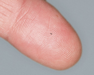
The Ex-PRESS™ Mini Glaucoma Shunt, a tiny 400-micron diameter stainless steel tube less than 3 mm long, lowers eye pressure by allowing aqueous humor to directly flow out of the eye. It is quicker and much less invasive when compared with traditional trabeculectomy surgery. Unlike trabeculectomy, it utilizes a very small incision using a specially designed disposable insertion tool, does not require partial removal of the iris (iridectomy), and has lower complications, faster recovery and minimal discomfort. Implantation of the device can reduce or eliminate the need for glaucoma eye drops. Bennett & Bloom Eye Centers doctors were among the first in the U.S. certified to perform this procedure. - XEN Gel Glaucoma Stent
The XEN Gel stent is one of the most recent advancements in glaucoma care and Bennett & Bloom is excited to be among the first in Kentucky to offer this technology to their patients. It is indicated for glaucoma that is either not responding or progressing despite drops. This tiny 6 mm device, about the width of a human hair, is inserted through a small corneal incision combined with regular cataract surgery or as a standalone procedure. The stent becomes soft and flexible providing long term decreased eye pressure. However, some patients may still require further eye drops to help control the pressure.
- Hydrus Microstent
This small flexible stent is roughly the size of a human eyelash and constructed of a nickel-titanium alloy. Like other minimally invasive surgeries, it is performed at the time of uncomplicated cataract surgery with minimal postoperative complications. The Hydrus microstent dilates or expands Schlemm’s canal, the eye’s natural drainage channel. The helps lower eye pressure and reduce the dependence on eye drops.
- Durysta
This biodegradable bimatoprost implant is compromised of the same active ingredient found in Lumigan, one of the most popular glaucoma drops. Durysta is painlessly inserted into the front part of the eye through a small injection in the office. It works similarly to Lumigan drops in that it enhances the eye’s natural drainage pathway to decrease intraocular pressure. Over 30% of eyes remain well controlled after 1 year and 25% at 2 years. Current FDA approval limits the eye to only a single injection.
.
- iStent Trabecular Micro-Bypass
- Surgery . Glaucoma surgeries are used as a last resort for patients not controlled with drops or laser. Postoperatively, patients are on strict activity restrictions for about a month and may have to miss work while the eye heals. Most have good eye pressure with minimal or no eye drops. However, glaucoma is not cured with surgery. Eye drops may need to be restarted and some may even require more surgery. It is very difficult to predict exactly how long a glaucoma surgery will last, with some working for over 10 years and others failing in under a year. The eye pressure still needs to be checked regularly. Surgery involves risks that less invasive lasers and procedures do not. Although rare, these risks can harm one’s vision. Rarely, the surgical site can develop a vision or eye threatening infection. If a glaucoma surgery patient ever develops a red, blurry, or painful eye, whether following recent surgery or many years later, they need to be examined right away to rule out infection.
- Trabeculectomy
Trabeculectomy surgery lowers eye pressure by allowing aqueous humor to directly flow out of the eye through a surgically created scleral incision. This procedure may be done alone, or in combination with cataract surgery if a visually significant cataract is present. - Drainage Device
These procedures lower eye pressure by allowing aqueous humor to flow directly out of the eye. Current implants include non-valved (Baerveldt, Molteno) and valved (Ahmed) devices. Drainage devices are less likely to get infections than Ex-PRESS™ or trabeculectomy surgeries. Drainage devices carry a very low risk of double vision that is typically treated with a change in glasses prescription.
- Trabeculectomy
- Eye drops
- Narrow-angle glaucoma
In most cases, narrow-angle glaucoma is treated by YAG laser iridotomy. The laser is used to create a painless opening in the iris (colored part of the eye) for fluid drainage. This may be preventative or may be performed in an emergency setting to prevent permanent vision loss and may need to be repeated depending on the iris color. Risks of bleeding, inflammation and a temporary increase in eye pressure are small. There are no postoperative restrictions. Your eye doctor typically prescribes an anti-inflammatory eye drop for a few days to help limit any post-laser irritation. In some instances, your doctor may recommend cataract surgery instead of a YAG laser iridotomy. Because artificial lenses are much thinner than the natural cataract lens, such replacement reduces the crowding that causes narrow angles in the first place. During cataract removal, the surgeon can sometimes break any scarring around the trabecular meshwork (goniosynechialysis) to improve its function and reduce dependence on pressure drops.
Will some medications worsen my glaucoma?
Many prescription and over-the-counter medications are labeled not to be used if you have glaucoma. These warnings apply only to untreated narrow-angle glaucoma. All systemic medications are safe with all other types of glaucoma.
Will I go blind from glaucoma?
Virtually no one should be blinded from glaucoma. Treatments are highly effective and, while vision already lost cannot be restored, the significant further loss can be avoided. The most important ingredient in successful glaucoma management is patient compliance. Continuous pressure control is possible only by strictly following your treatment plan…and that is primarily up to you. In most cases, the only people who become blind from glaucoma are those who do so before their condition is diagnosed or who do not follow their doctor’s advice and treatment regimen.


