How are tears produced and drained?
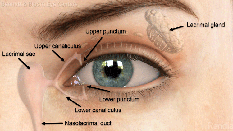
Tears are produced by the lacrimal gland. The tears then pass over the eye surface and drain through the upper and lower puncti, the upper and lower canaliculi, and then into the lacrimal sac which is located near the side of the bridge of the nose. Finally, the tears pass through the nasolacrimal duct which drains into the nasal cavity and into the back of the throat. This is why you can often taste tears or eye drops.

What are the symptoms of a blocked tear duct (nasolacrimal duct obstruction)?
Patients may experience excessive tearing (epiphora). This may be constant or intermittent and may cause surrounding redness and skin tenderness. This is often associated with mucus discharge and mild conjunctivitis. In some cases, an infection can develop underneath the skin between the eye and the nose (dacryocystitis).
What causes a blocked tear duct ?
Infants frequently develop nasolacrimal duct obstruction due to incomplete development of the drainage system and the nasal cavity. Adults may develop this at any time due to normal age-related changes, trauma, inflammation, infection, or mechanical obstruction.
How is nasolacrimal duct obstruction treated?
In infants, the nasolacrimal duct obstruction often resolves spontaneously but may also be relieved by digital massaging as instructed by your eye doctor. For those that don’t resolve, minor outpatient surgery is usually successful.
For those with persistent tearing without an infection, nasolacrimal duct probing with stent insertion is usually recommended. This is performed either in the office or in the operating room under local anesthesia. Increasing sized probes are inserted in the normal tear passageway until it is fully dilated. A soft plastic tube is placed during surgery to prevent the passage from becoming blocked again. This tube is removed in the office after 6 to 9 weeks.
For those with secondary infections or if probing fails to correct the problem, dacryocystorhinostomy (DCR) is needed. The DCR by-passes the nasolacrimal duct to create a new passage for the tears to drain. Under general anesthesia, a small opening in the nasal bone is created with either a small skin incision or internally through the nose. A tube is placed in the new passage to keep it open. The tube is then tied and stitched to the inside of the nose. The tube is removed in the office about 6 months postoperatively.
What are the major risks of nasolacrimal duct obstruction surgery?
Risks include infection, scarring, and spontaneous extrusion of the tube. In less than 10% of cases the new drainage channel may not stay open, which may require additional surgery.
What are the alternatives to surgery?
You may decide to live with the tearing, discharge, and irritation that a blocked tear duct can cause. However, if you have had an infection, your surgeon will likely recommend surgery since recurrent infection can rarely cause vision loss.
If your tear duct is partially blocked, a balloon can be inflated and/or tubes placed to enlarge the duct and keep it open. However, if your tear duct is completely blocked, there are no other options besides the above procedures.


