What is a choroidal rupture?
The macula is the small, specialized area of the retina that gives us our straight-ahead reading and driving vision. Blunt eye trauma temporarily causes the round eye to stretch out like a football. This can stretch the macula and tear the orange eye wall, causing a choroidal rupture.

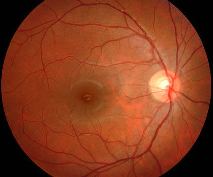
Normal macula

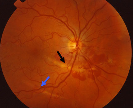
Two choroidal ruptures following recent blunt trauma. One of these (black arrow) has some associated blood.
How is a choroidal rupture diagnosed?
A choroidal rupture is a clinical diagnosis found during a dilated retinal examination. Immediately following the blunt injury, blood can hide the underlying rupture. Fluorescein angiography is often used to confirm the diagnosis and assess the severity of the retinal damage. Once the blood clears it is much easier to tell the extent of the macular damage. Central vision is usually good unless the rupture extends through the macular center.
New blood vessel growth beneath the macula (macular neovascularization, MNV) can develop in some patients with choroidal ruptures. These vessels cause the macula to swell with fluid and blood and often lead to permanent central vision loss. There are many causes of CNV including angioid streaks, degenerative myopia, idiopathic, ocular histoplasmosis, and age-related macular degeneration.
How is a choroidal rupture treated?
There is no specific treatment for the choroidal rupture, although CNVM can be treated with Avastin, Lucentis or Eylea. These drugs belong to a new class of potent medications, VEGF inhibitors, that prevent CNVM from growing and leaking. They have been extensively studied in patients with age-related macular degeneration, and are also highly effective in choroidal ruptures. Click here to learn more.
View more retina images at Retina Rocks, the world’s largest online multimedia retina image library and bibliography repository.


