What is Coats’ Disease?
The retinal blood vessels work like a garden hose, bringing oxygen and other nutrients into and out of the eye. In Coats’ disease, these vessels are weak, allowing fluid, blood, and lipid (fatty deposits) to leak into and beneath the retina causing it to swell and not work properly. Central and peripheral vision can become blurred, just as a water droplet placed on a photograph will cause the picture to blister and become distorted.
Very mild cases can be totally asymptomatic. Severe leakage can cause extensive vision loss from lipid and fluid damaging the macula or from retinal detachment.
Coats’ disease predominantly occurs in males and only affects one eye. It is not associated with any other eye or systemic abnormalities.

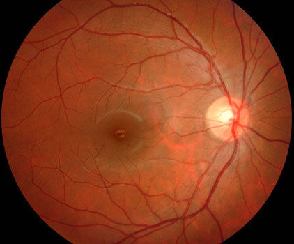
Normal macula

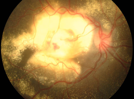
Coats’ disease with severe accumulation of yellow lipid under the macula.
How is Coats’ Disease diagnosed?
Coats’disease is a clinical diagnosis found during a dilated retinal examination. Fluorescein angiography is often used to confirm the diagnosis and assess the severity of the retinal damage. The more severe disease generally presents earlier in life with painless vision loss. In young children, parents may notice the affected eye wandering. Photographs of the child may also show a yellow discoloration of the pupil.
How is Coats’ Disease treated?
The leaking blood vessels are treated with laser photocoagulation or cryotherapy. Treatment often needs to be repeated multiple times to control pre-existing leaks or for new lesions.

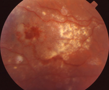
Extensive retinal hemorrhages and fatty deposits before treatment.

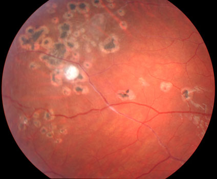
The exudation resolved following several laser treatments.
View more retina images at Retina Rocks, the world’s largest online multimedia retina image library and bibliography repository.


