What is degenerative myopia?
The normal eye is a round ball about 24 mm (0.94 inches) in diameter. Far-sightedness (hyperopia) occurs when the eye is shorter than normal and near-sightedness (myopia) when the eye is larger. In degenerative myopia the eye becomes elongated. The retinal and orange choroidal tissues that line the eye become thinned. There can also be frank out-pouchings (posterior staphyloma) of the macula.
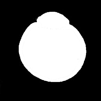
Normal eyeball
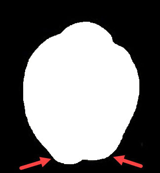
Elongated eyeball with posterior staphylomas (arrows)
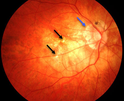
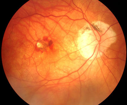
The orange macular pigment is thinned and yellow. Small breaks in the orange macular pigment are seen (lacquer cracks, black arrows). There is also thinning around the optic nerve (blue arrow).
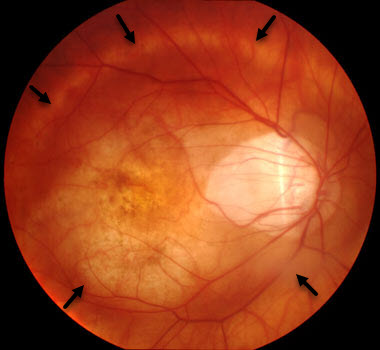
Out-pouching of the eyewall (posterior staphyloma)
Some patients can develop splitting of the retinal tissue (macular retinoschisis) due to macular pucker, vitreomacular traction, or to the macular tissue being stretched by the elongation of the eye. This can cause central vision loss and distortion.
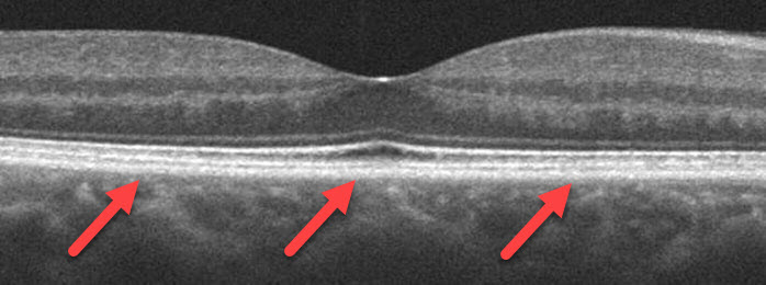
OCT scan of a normal macula. The arrows mark the back of the retina which is flat against the eyewall.
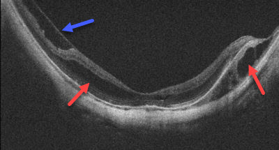
The eyewall and retina are bowed posteriorly with retinoschisis (red arrows) and a macular pucker (blue arrow)
New blood vessel growth beneath the macula (macular neovascularization, MNV) can develop in some patients with degenerative myopia. These vessels cause the macula to swell with fluid and blood that can lead to permanent central vision loss. There are many causes of MNV including angioid streaks, choroidal rupture, idiopathic, ocular histoplasmosis, and age-related macular degeneration. Fluorescein angiography and OCT scanning help diagnosis the presence of MNV.
Patients with degenerative myopia develop floaters and posterior vitreous detachment earlier in life. They are also at risk for retinal breaks and detachment.
What treatments are available for degenerative myopia?
Eliminate the need for glasses and contact lenses.
LASIK and vision correction surgeries offer many exciting options to eliminate the need for glasses and contact. Click here to learn more about these procedures.
Macular neovascularization.
Avastin, Lucentis, and Eylea belong to a new class of potent medications, VEGF inhibitors, that prevent MNV from growing and leaking. They have been extensively studied in patients with age-related macular degeneration, and are also highly effective in myopic MNV. Click here to learn more.
Macular retinoschisis.
The vast majority of patients with macular retinoschisis maintain excellent vision and don’t require any treatment. Some will develop central vision loss and distortion. Vitrectomy surgery, similar to that done for eyes with macular pucker or vitreomacular traction, are often successful.
Retinal breaks and detachment.
Increasing amounts of nearsightedness increase the risks for retinal breaks and detachment. Click here to learn more about how these are treated.
View more retina images at Retina Rocks, the world’s largest online multimedia retina image library and bibliography repository.


