What is X-linked retinoschisis?
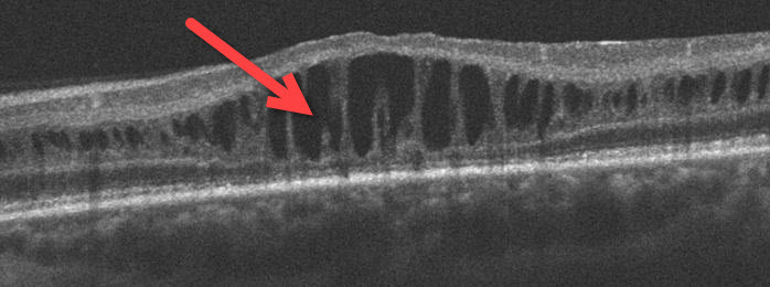
The retina lines the inside of the eye like wallpaper. It has multiple layers that are normally stuck together. Sex-linked retinoschisis is a genetic disorder where the retinal layers split. Splitting can occur in the macula or in the peripheral retina.

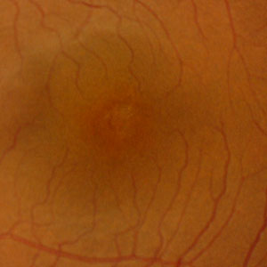
Normal retina

X-linked retinoschisis with macular splitting
Sex-linked retinoschisis usually affects males due to an abnormality on the X chromosome. All males exhibit the retinal findings and all female carriers are unaffected. An affected male transmits the disorder to half of his grandsons through each of his daughters.
How is X-linked retinoschisis diagnosed?
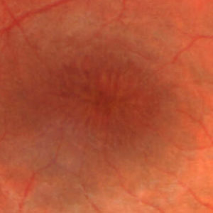
Patients usually notice central vision loss or floaters in childhood. The retinal changes, which affect both eyes, are detected during a dilated retinal examination and with OCT scanning. The central macula has radiating spokes of the split retina. The peripheral retina can also have elevated sheets of retinal splitting.

Normal macula

X-linked retinoschisis with radiating spokes of retinal splitting
How is X-linked retinoschisis treated?
There is no specific treatment for the underlying genetic defect. Most of the retinal changes occur within the first few decades of life, and the vision usually levels off between 20/50 and 20/100. Most patients maintain a normal active life.
Carbonic anhydrase inhibitor eye drops (dorzolamide or brinzolamide) are sometimes used to minimize the macular retinoschisis. Vitrectomy surgery is rarely necessary to remove non-clearing vitreous hemorrhage or for retinal detachment.
View more retina images at Retina Rocks, the world’s largest online multimedia retina image library and bibliography repository.


