What is Pseudotumor Cerebri (PTC)?
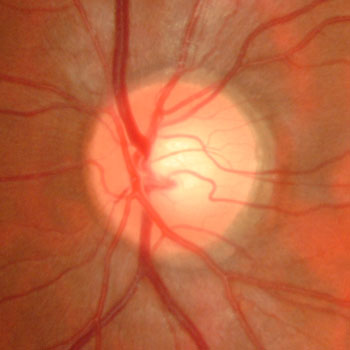
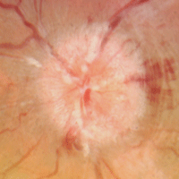
The images formed by the retina (the eye’s “film”) are sent to the brain thru the optic nerve. As it exits the back of the eye, the optic nerve appears as a distinct yellow-pink circle. There are various serious brain conditions, including tumors, strokes, infection, and bleeding that can cause the nerve to swell.

Normal optic nerve

Swollen optic nerve
The brain, encased in the bony skull, is cushioned by the cerebrospinal fluid (CSF). Chronically elevated CSF pressure can cause the optic nerve to swell. When this happens for no other identifiable cause, this is called pseudotumor cerebri (PTC) or idiopathic intracranial hypertension. Although the underlying cause is unknown, PTC is more common in younger obese women or with certain medications (birth control pills, steroids or tetracycline). About 10% of cases are caused by cerebral venous stenosis or thrombosis.
What are the symptoms of PTC?
The most common symptoms include headaches and blurred vision (especially when bending over). Untreated PTC can cause severe vision loss.
How is PTC diagnosed?
PTC is usually suspected when a patient presents with bilateral optic nerve swelling (papilledema) found during a dilated eye examination. An MRI scan is ordered to rule out other causes of papilledema (including a tumor), and an MRV scan is also performed to look for cerebral venous abnormalities. If the MRI scan is normal, a lumbar puncture (spinal tap) is performed. Elevated CSF pressure confirms the diagnosis of PTC. OCT scanning can precisely measure the amount of optic nerve swelling, and visual field testing documents peripheral vision loss.
How is PTC treated?
PTC is usually managed with a team of health care professionals including your eye doctor and a neurologist. Causative medications are stopped and weight loss is recommended. CSF fluid removed during the diagnostic spinal tap can sometimes relieve the elevated CSF pressure and symptoms. Patients are usually placed on oral acetazolamide (Diamox), a diuretic. Treatment is often necessary for many months or years. Rarely surgical procedures (optic nerve sheath fenestration, cerebrospinal fluid shunting, or intracranial venous sinus stenting) are necessary.
As the CSF pressure normalizes, the optic nerve swelling, headaches, central and peripheral vision loss will usually improve. Patients are followed regularly by their eye doctor who literally functions as the “eyes” for the neurologist, letting them know if the ocular findings are responding to treatment.
View more retina images at Retina Rocks, the world’s largest online multimedia retina image library and bibliography repository.


